carcinoma breast case presentation .pptx
This document summarizes the case of a 62-year-old female patient who presented with a lump in her right breast. On examination, a 4 cm irregular, mobile lump was detected. Investigations including mammography and biopsy confirmed a diagnosis of invasive ductal carcinoma. The patient underwent a modified radical mastectomy with axillary clearance. Histopathology of the specimen found grade 2 invasive ductal carcinoma with clear margins and no lymph node metastasis. The final diagnosis was invasive ductal carcinoma of the right breast, stage T2N0M0. Read less


More Related Content
- 1. Welcome to Clinical Case Presentation Dr. Md. Tafazzul Hossain Bhuiyan IMO SU III DMCH
- 2. A 62 Year Old Female With Breast Lump
- 3. Particulars of the patient • Name: Johura Begum • Age: 62 years • Sex: Female • Address: Shibaloy, Manikgonj • Marital status: Married • Occupation: Housewife • Religion: Islam • Date of admission: 19/09/2022 at 3.30pm • Date of examination: 19/09/2022 at 4.00pm
- 4. Present complaint • Lump in the right breast for 1 month
- 5. History of the present complaint According to the patient’s statement she was relatively well about 1 month back, then she suddenly noticed a lump in upper part of right breast which was gradually increasing in size. She had no pain and fever. She had no history of trauma to the breast. She had no complaint of bone pain, cough, chest pain or weight loss. Her bowel and bladder habit was normal. She did not give history of diabetes, hypertension or contact with TB patient.
- 6. Past medical history • She had no significant past medical or surgical illness
- 7. Drug history • She did not took any medication for this disease
- 8. Family history • Her mother died naturally at old age • She has 2 sisters, none has this type of illness • 1 daughter is in healthy state
- 9. Personal history • She is non smoker, non alcoholic, occasional betel leaf and betel nut chewer Socio economic history • Middle socio economic condition • Lives in brick built house with tin shed roof, use sanitary latrine, drinks safe water from tube well
- 10. Immunization history • She took BCG vaccine at young age • She is vaccinated against COVID19
- 11. Allergic history • She had no history of allergy to any known medication or food. Obstetric history • Married for 50 years • Para 3(NVD) • Menarche at 12 years • Menopause at 40 years
- 12. Breast feeding history • She breast fed her 2 sons and 1 daughter Contraceptive history • She was use to take contraceptive pill irregularly
- 13. General examination • Appearance: normal • Body build: normal • Cooperation: co-operative • Decubitus: on choice • Nutritional status: average(BMI 25.4) • Anemia: absent • Cyanosis: absent • Jaundice: absent • Edema: absent
- 14. General examination (cont) • Dehydration: absent • Clubbing: absent • Koilonychia: absent • Leukonychia: absent • Pulse: 96bpm • BP: 130/80 mmHg • Respiratory rate: 18bpm • Temperature: 98⁰F
- 15. General examination (cont) • Lymph nodes: accessible nodes are not palpable • Thyroid: not enlarged • Skin condition: normal • Bony tenderness: absent
- 16. Local examination Inspection • Both breasts are normal in size and shape • Nipples are normal and symmetrical • No visible lump • No ulcer or peau d’orange or skin tethering • No scar mark, engorged vein • No discharge from nipples
- 17. Inspection • Picture was taken with proper consent of the patient
- 18. Palpation • Left breast: normal and no palpable lump • Right breast: There is a lump in upper and outer quadrant Tenderness: no tenderness Temperature: no local rise of temperature Consistency: hard
- 19. Palpation (cont) Shape: globular Margin: irregular Size: about 4 cm in its maximum diameter Fixity: mobile in all direction and free from underlying structure and overlying skin • Axilla: no palpable nodes in any axilla
- 20. Abdominal examination Inspection • Skin normal • Flanks full • Umbilicus centrally inverted • No scar mark • No visible peristalsis • No engorged veins Palpation • Superficial palpation • Temp: normal • Tenderness: absent • Deep palpation • Lump: no lump felt • Liver: not enlarged • spleen: not enlarged • Kidneys: non ballotable
- 21. Abdominal examination (cont) Percussion • Percussion note: tympanitic • Liver dullness: right 5th intercostal space in mid clavicular line • Shifting dullness: absent Auscultation • Bowel sound: present • Hepatic bruit: absent • Renal bruit: absent
- 22. Other systemic examination Respiratory system • Inspection: normal findings • Palpation: no abnormality seen • Percussion: resonant • Auscultation: breath sound normal, no added sound Cardiovascular system • Inspection: normal findings • Palpation: no cardiomegaly, no palpable thrill or murmur • Auscultation: normal heart sound, no murmur heard
- 23. Other systemic examination (cont.) • Musculoskeletal system: no abnormality or any bony tenderness found • Nervous system: normal • Others systems are apparently normal
- 24. Salient feature Mrs. Johura begum, a 62 years postmenopausal, normotensive, nondiabetic lady was presented with a painless hard lump in upper and outer quadrant of right breast for 1 month. She had no positive family history of any malignant diseases. She had no history of trauma to the breast and no bone pain.
- 25. Salient feature (cont) On examination the lump was about 4 cm in its maximum diameter, margin was irregular, mobile, non tender and no local rise of temperature. There were no skin changes over the lump. There was no nipple discharge. Left breast was normal and there was no axillary lymphadenopathy. Her all vital parameters were normal. Other systemic examination were normal.
- 26. Provisional diagnosis
- 27. Carcinoma right breast (T2N0Mx)
- 28. Differential diagnoses • Phyllodes tumor • Traumatic fat necrosis
- 29. Investigations • Mammography : right breast is predominantly fatty. A radio opaque shadow is seen in upper and inner quadrant of right breast. No micro or macro calcification is noted. Overlying skin and soft tissue appears normal. • Impression: suspicious mass in right breast. Right axillary lymphadenopathy • Category: BIRADS 4
- 30. Mammography
- 31. USG of both breast and axilla • A fairly solid mass lesion with lobulated margin is noted in right breast at 12o’ clock position • No abnormal micro or macro calcifications could be noted • A lymph node <1cm demonstrated in right axillary region • Impression: Right upper mid quadrant (12o’ clock) solid mass with enlarged right axillary lymph node most likely malignant
- 32. USG of both breast and axilla
- 33. Core biopsy and histopathology • Gross: specimen consists of 3 linear pieces of tissues. • Microscopic examination: section shows core of breast tissue. It reveals an invasive ductal carcinoma. The tumor shows mild desmoplastic changes. Nottingham histologic score: 6 • Histologic grade II • Lymphovascular invasion: not identified • Perineural invasion: absent • Diagnosis: Invasive ductal carcinoma, NOS, grade II
- 34. Immunohistochemistry • Estrogen receptor (ER): positive • Progesterone receptor(PR): positive • HER-2: negative • Ki67: immunoreactive in 12% tumor cells
- 35. Histopathology & immunohistochemistry
- 36. Investigations Investigations Date Reports CBC 19/09/22 Hb- 14.1gm/dl, WBC- 12,490/cmm, PLT-216*10^3/mm3 S. glucose 19/09/22 7.2 mmol/l S. creatinine 19/09/22 1.24 mg/dl S. ALT 24/09/22 41U/L S. electrolytes 19/09/22 Na+ 143, K+ 4.6, Cl- 106 mmol/L Blood grouping Rh typing 19/09/22 A+ve ECG 19/09/22 Normal HBsAg and Anti HCV 19/09/22 Negative CXR PA view 19/09/22 Normal study USG of WA 26/09/22 Fatty liver
- 37. Clinical diagnosis • Invasive ductal cell carcinoma (right breast) grade II, NOS (T2N0M0)
- 38. Management • Counselling • Preoperative assessment • Multidisciplinary approach • Operation of the patient • Adjuvant chemotherapy • Rehabilitation and psychological support
- 39. Operation note • Date and time: 28/09/22 at 1.50pm • Name of operation: modified radical mastectomy with axillary clearance of right breast • Indication: Carcinoma of right breast with rt axillary lymphadenopathy • Incision: transverse elliptical incision • Anesthesia: GA
- 40. Operation note (cont) • Findings: a lump present in upper and outer quadrant, right axillary lymphadenopathy • Procedure: with all aseptic precaution proper painting and draping was done. Incision was given. Modified radical mastectomy was done. Axillary clearance was done. Two negative suction drain was kept. Skin was closed. • Specimen sent for histopathology
- 41. • Patient was discharged in 3rd post operative day • With advice to consult with dept of radiotherapy with histopathology report
- 42. Histopathology of specimen • Invasive ductal carcinoma • Grade 2( Nottingham modification of Bloom Richardson system) • Lymphovascular invasion: not identified • Perineural invasion: not identified • Tumor extension: skin: free of tumor • Nipple and areola: free of tumor • Other quadrants: free of tumor
- 43. Histopathology (cont.) • Resection margin: deep resection margin clear • Peripheral resection margin clear • Lymph nodes(17): reactive hyperplasia, no metastasis seen • Pathological staging: pT2N0Mx • Diagnosis: Invasive ductal carcinoma, NST grade 2
- 44. Final histopathology report
- 45. Final diagnosis • Invasive ductal carcinoma (right breast), Grade II, NOS (T2N0M0)
- 46. Thank you
Case Study 63: Cancer of the Female Breast
Dec 21, 2019
170 likes | 259 Views
Case Study 63: Cancer of the Female Breast. By Robyn Schwartz. Case Background. 46, premenopausal Dense breasts Has noticed cysts in the past Noticed new lump in upper right quadrant Did not resolve Got bigger Denied lumps in axillary. Patient history. Happily married for 21 years
Share Presentation

Presentation Transcript
Case Study 63: Cancer of the Female Breast By Robyn Schwartz
Case Background • 46, premenopausal • Dense breasts • Has noticed cysts in the past • Noticed new lump in upper right quadrant • Did not resolve • Got bigger • Denied lumps in axillary
Patient history • Happily married for 21 years • 3 kids (3, 8, and 10) • Does breast self exams • Normal pap 2 years ago • Has Asthma and hypertension • Exercises • No tobacco, alcohol or illegal drugs
Risk Factors • All 3 kids born after the age of 35 • First period at 11yr old • Dense breasts • Cysts already develop regularly • Family history of breast cancer • Paternal grandmother diagnosed at age 45 before menopause • Mother diagnosed at age 45 before menopause. Died at age 73 from reoccurrence of breast cancer
Breast Cancer: What is it? • Uncontrolled division of abnormal cells in the breast • Caused by specific mutations • BRCA1 and BRCA2 • TP53
Our patient: Mammogram • 2.3cm x 2.9cm x 3.2cm mass • Irregular borders • Skin thickening • Enlarged axillary lymph node • 6 Y-shaped microcalcifications extended toward nipple • Abnormal mass into pectoral muscle
Grading vs Staging • How far the cancer has spread • I, II, III, IV • Based on • Size of tumor • Invasive vs non invasive • Spread to lymph nodes • Spread to other parts of the body • How abnormal the cells are • 1, 2, 3,4 • Based on • Tubule formation • Size and shape of cells • Mitotic division • Measures the likely aggressiveness of the cells
Grading tumors Nuclear (size/shape) Score 1: little variation in size Score 2: moderate variability, open vesicular nuclei Score 3: lots of variability open nuclei Mitotic Score 1: <7 mitoses Score 2: 8-14 mitoses Score 3: >14 mitoses Tubular differentiation Score 1: > 75% glandular/tubular Score 2: 10-75% glandular/tubular Score 3: < 10% Glandular/ tubular
Staging Cancer • agrdjdytydstasf Stage 0: No Cancer Stage I: IA: Cancer is small, low grade and localized IB: Cancer is large, low grade and localized Stage II: IIA: Tumor is 2-5cm but has not spread IIB: Tumor is 2-5cm but has spread to lymph nodes Stage III: Tumor is larger than 5cm and has spread to multiple lymph nodes Stage IV: Cancer has spread to other parts of the body
Our Patient: Biopsy and ultrasound • Ultrasound: • Non-cystic mass, solid appearing • Abnormal vascularity • Some skin thickening and mild tissue edema • Biopsy: • Consistent with infiltrating breast cancer • 3-5 divisions per high power field • Mild pleomorphism • Positive for estrogen and progesterone receptors
Grade and Stage • Grade 1 • Mitotic score: 1 (<7 divisions) • Glandular Score: 1 (75% glandular) • Nucleic Score: 1 (not much change) • Total score: 3 • 10 year survival rate 90% • Stage IIB • Small • Spread to 1 lymph node • 5 year survival rate of 71%
Our Patient: Treatment • Breast conservation therapy • Lump removal • Radiation • Lymph node biopsy • Tamoxifen • Estrogen receptor blocker • Helps stop growth of cancer cells
Our Patient: Follow Up • 6.5 years cancer free • 80 months later, complained of • bone pain in lower back • Headache
Test Results • Bone scan • Lesions in lumbar spine without fracture • Chest X-Ray • 3 small nodules in upper lobe of left lung • Brain MRI • Small mass in right frontal lobe • Abdominal CT • Negative • Blood tests • CEA elevated by 2-fold • CA27-29 concentration elevated by 2-fold
Diagnosis, Outlook, and Treatment • Stage IV Breast cancer • 13% 10-year survival rate • Treatments • Chemotherapy • taxanes • Hormone Therapy • Targeted therapy • HER2 targeted therapy • Slow growth • Manage Bone Metastasis • Biophosphonates • Slow destruction
How to Prevent cancer • Exercise • Eat well • Don’t smoke • Do regular breast self exams • Report anything suspicious immediately • Check family history
- More by User

Breast Cancer, BRCA2 Gene
Breast Cancer, BRCA2 Gene Breast Cancer Breast cancer specifically refers to a cancer that forms in tissues of the breast Usually in the ducts – which are the tubes that carry milk to the nipple Or the lobules – glands that make milk It occurs in both men and women
1.5k views • 39 slides

BREAST CANCER STUDY
BREAST CANCER STUDY A STUDY OF ISSUES INVOLVING THE DIAGNOSIS OF BREAST CANCER THAT RESULT IN MEDICAL MALPRACTICE CLAIMS 2002 Breast Cancer Study Focus 450 cases involving paid claims with resolution dates no earlier than January 1, 1995 were analyzed.
774 views • 32 slides
ACP: Breast Cancer Update
ACP: Breast Cancer Update. Elaine A. Muchmore, M.D. UC San Diego 10/20/07. Breast Cancer. Breast cancer affects more than 150,000 women each year, with a lifetime incidence of greater than 10%.
653 views • 19 slides

Breast cancer screening
[email protected]. 2. . Breast cancer screening. Early detection is an important factor in the success of breast cancer treatment. The earlier breast cancer is found, the more easily and successfully it can be treated. The three methods commonly used for early detection are: Breast self-exam (BSE). Clinical breast exam (CBE).Mammogram..
1.45k views • 32 slides

Ambi Journal Club June 14th 2006 Breast Cancer
ACADEMIC APPROACH TO BREAST CANCER IN LITERATUREDrug Therapy: Treatment of Breast Cancer Hortobagyi G. N. N Engl J Med 1998; 339:974-984, Oct 1, 1998. Review ArticlesPrimary Care: Assessing the Risk of Breast Cancer Armstrong K., Eisen A., Weber B.
942 views • 49 slides

THE REGISTRATION OF BREAST CANCER
2. FIVE FACTS ABOUT BREAST CANCER. Incidence:1 in 9 women in the TCR area develop breast cancer during their lifetime.0.5% of breast cancer cases occur in men.Survival:Now improving. Depends on stage at diagnosis.90% at 5yrs for localised disease, 77% at 5 yrs if lymph nodes involved.Most
435 views • 24 slides

Breast & Prostate Cancer
Breast & Prostate Cancer. Wednesday, October 17 2007. Introduction. Who we are Why we’re here today What is breast cancer What is prostate cancer Statistics How this affects you Prevention or Living with it. Breast Cancer. What is Breast Cancer?.
784 views • 31 slides
Breast Cancer
Breast Cancer. Kristi McIntyre M.D. Resident’s lecture. Breast cancer. Risk factors Hereditary breast cancer Detection Staging Surgical intervention Prognostic factors Management of early stage breast cancer. Breast cancer risk factors. Age/race
1.55k views • 59 slides

Weight Control for Breast Cancer Prevention
Outline. Weight control and breast cancer: BackgroundEvidence for primary breast cancer riskEvidence for breast cancer survivalPlanning of a national trial in weight control2. Practical aspects of weight control3. Proposed pilot study of weight control in newly diagnosed women though
905 views • 79 slides

Cancer Breast
Cancer Breast . * Epidemiology:. The most common malignant tumor in female. Accounts for 32% of all female cancer.
469 views • 18 slides
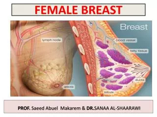

FEMALE BREAST
FEMALE BREAST. PROF. Saeed Abuel Makarem & DR. SANAA AL-SHAARAWI. OBJECTIVES. By the end of the lecture, the student should be able to : Describe the shape and position of the female breast. Describe the structure of the mammary gland. List the blood s upply of the female breast.
1.39k views • 17 slides
R5 洪逸平 SUPERVISOR 趙大中醫師
R5 洪逸平 SUPERVISOR 趙大中醫師. Breast Cancer. The most prevalent cancer in female Mortality 4 th in Taiwan. Treatment of Breast Cancer. Breast Cancer. When to change regimen? Unacceptable toxicity Progression disease. Current Tools for Follow-up. Radiologic image Standard serologic test
419 views • 27 slides

Breast cancer
Breast cancer. PPT 22 nd November 2012 Introduction: Dr B K Jindal Mr A Haq Consultant Surgeon . Aims and Objectives: . To understand the prevalence of breast cancer To receive the trends in survivability from breast cancer To consider the outcome of 2 week wait referrals
812 views • 26 slides

Physical Activity & Breast Cancer
Physical Activity & Breast Cancer. By Janet Foote, PhD. Educational Objectives. Discuss the role of movement in: 1. Prevention of breast cancer onset 2. Appropriateness for breast cancer survivors 3. Prevention of Breast Cancer recurrence.
525 views • 25 slides

Neoadjuvante Therapy of Breast Cancer
Neoadjuvante Therapy of Breast Cancer. Cancer statistics Genesis of cancer Cancer in detail Treatment of cancer Prevention Follow-up NEWS Research. INCIDENCE OF CANCER. Colorectal cancer Breast cancer Lung Cancer Prostate cancer Cervix carcinoma Meaning of
1.06k views • 55 slides

Breast Cancer. Kathrina Calulut Alison Saechao. Breast Cancer. Cancer of tissues of the breast Ductal carcinoma Lobular carcinoma. Risk Factors. 1 in 8 women Age and Gender Family History Substance Abuse Childbirth Obesity. Early Symptoms. New lump or mass
817 views • 15 slides
Development of laboratory models to study Breast Cancer
Section of Pathology and Tumour Biology. Development of laboratory models to study Breast Cancer. Deborah Holliday Breast Research Group Section of Pathology & Tumour Biology. Outline. Introduction to the cells found in breast tissue Changes in breast cells during breast cancer
376 views • 20 slides

HER-2 Positive Breast Cancer Market in the US 2015-2019
Breast cancer is characterized by uncontrolled growth of cancerous cells in the breast. It occurs in both males and females, however, the incidence of breast cancer in males is rare. Histologically, breast cancer can be classified into ductal carcinoma, lobular carcinoma, nipple cancer, and other undifferentiated carcinoma. Breast cancer is the second most common form of cancer in women. HER-2 is a protein that affects the growth of malignant cells and is usually found on the surface of normal breast cells. Sometimes, a large number of HER-2 receptors are found on the surface of breast cancer cells. Get full report & TOC @: http://www.researchbeam.com/her-2-positive-breast-cancer-in-the-us-2015-2019-market
314 views • 6 slides

Latest Study on Global Breast Biopsy Devices Consumption with Investment Feasibility Analysis and Market Research on Maj
A breast biopsy is a procedure in which part or all of a suspicious area in the breast is removed and examined, usually for the presence of cancer. Breast cancer is the most common cancer affecting women and accounts for the second highest incidence of cancer-related death, after lung cancer.
285 views • 7 slides

Breast Cancer Therapeutics in Asia-Pacific Markets to 2021
Breast cancer, a malignant neoplasm, is the second-most common cancer and the most common cancer in women worldwide, accounting for 16% of all female cancers, making the disease exceedingly prevalent. For More Information: http://bit.ly/1NnH1v0
289 views • 8 slides

How Breast Cancer Prevent With Cancer Treatment Centers?
If cells of breast spread out of control and forms a tumor it is the sign of Breast cancer with the help of X-ray we seen this changes. Many Cancer Treatment Centers are providing awareness for breast cancer because in this case, less symptoms are happening. There are 4 stages of breast cancer it can depend on changes in the breast. Houston Cancer Specialist are offered many therapies to stop breast cancer and also get success. If any treatment of Houston cancer hospital does not give the best result of treatment then also take the second opinion for cancer sometimes it shows best result.
747 views • 11 slides

Breast Cancer : Overview of symptoms, causes, diagnosis, risk factor and treatment
Breast cancer is a disorder in women, which starts in the inner lining of milk ducts or the lobules that supply them milk. Breast cancer may include lump in the breast, a change in breast or red scaly patch on skin. Breast cancer usually builds up with the age or it can be genetics.
506 views • 14 slides

- My presentations
Auth with social network:
Download presentation
We think you have liked this presentation. If you wish to download it, please recommend it to your friends in any social system. Share buttons are a little bit lower. Thank you!
Presentation is loading. Please wait.
To view this video please enable JavaScript, and consider upgrading to a web browser that supports HTML5 video
Case Study 63: Cancer of the Female Breast
Published by Lilian Paul Modified over 9 years ago
Similar presentations
Presentation on theme: "Case Study 63: Cancer of the Female Breast"— Presentation transcript:
Cancer -uncontrollable or abnormal growth of abnormal cells. *1st leading cause of death is a heart attack *Cancer is the 2nd leading cause of death.

What is cancer? A cancer is a malignant tumor, which are cells that multiply out of control, destroying healthy tissues (Dictionary)

Breast Cancer 101 Barbara Lee Bass, MD, FACS Professor of Surgery

Breast Cancer Prevention & Early Detection

Breast Cancer Liz Ignatious, Maddie Ticer, Molly Houlahan.

Muamer Martincs Shontez Miller Sokol Talovic. She's 35 years old white female Lives in Massachusetts, CT She has a strong family history of Breast.

Breast Cancer Nick Settecase, Payton Picone, & Mike Malone.

Breast Cancer Kathrina Calulut Alison Saechao. Breast Cancer Cancer of tissues of the breast Ductal carcinoma Lobular carcinoma.

By Rachel, Xiao Xia, Helen. Introduction Definition Symptoms Causes Prevention Treatment Prognosis Statistics Conclusion.

Do Now #4 What is cancer? What are some warning signs of cancer? What are some forms of treatment?

Breast Cancer By: Vincent Russo And Scott Jeffery.

Reproductive health. Cancer Definition Cancer Definition The abnormal growth of cells without normal control of body. Types of Cancer Malignant Cancer.

Breast Cancer Presentation by Dr Mafunga. Breast cancer in the UK Breast cancer is the second most common cancer in women. Around 1 in 9 women will develop.

Breast Cancer By George Rezk.

Breast Cancer This slide goes first.

BREAST CANCER AWARENESS Sheraton Kuwait , Crystal Ballroom

BREAST CANCER GROUP 6 : Nuraini Ikqtiarzune Haryono( ) Tri Wahyu Ningsih ( ) Rani Yuswandaru ( ) Anita Rheza Fitriana Putri( )

Breast Cancer Awareness By: Dominick Phillips. What Is Breast Cancer? If a cell changes into a abnormal, sometimes harmful form, it can divide quickly.

عمل الطالبات : اسماء جادالله فاطمة الحشاش ختام الكفارنة.
About project
© 2025 SlidePlayer.com Inc. All rights reserved.

IMAGES
COMMENTS
Jan 29, 2013 · This case study describes a 37-year-old female patient who presented with a breast mass. Diagnostic tests performed included a mammogram, biopsy, and right modified radical mastectomy which revealed invasive ductal carcinoma.
Dec 23, 2019 · This case study summarizes a 38-year old female patient presenting with breast pain, nipple tenderness, and bloody discharge. Her medical history includes a diagnosis of tennis elbow. Laboratory tests and cytology reports were conducted.
Feb 3, 2024 · This document summarizes the case of a 62-year-old female patient who presented with a lump in her right breast. On examination, a 4 cm irregular, mobile lump was detected. Investigations including mammography and biopsy confirmed a diagnosis of invasive ductal carcinoma.
Oct 1, 2014 · Case Study: Radiation Therapy and Ultrasound Management of Breast Cancer. hhholdorf. Radiation Therapy (also known as radiotherapy and radiation oncology) began shortly after the discovery of X-rays in 1895 by Wilhelm Rontgen.
Nov 13, 2014 · Case Study 63: Cancer of the Female Breast By Robyn Schwartz. Case Background • 46, premenopausal • Dense breasts • Has noticed cysts in the past • Noticed new lump in upper right quadrant • Did not resolve • Got bigger • Denied lumps in axillary
Dec 21, 2019 · Breast Cancer : Overview of symptoms, causes, diagnosis, risk factor and treatment. Breast cancer is a disorder in women, which starts in the inner lining of milk ducts or the lobules that supply them milk. Breast cancer may include lump in the breast, a change in breast or red scaly patch on skin.
Breast Cancer Awareness By: Dominick Phillips. What Is Breast Cancer? If a cell changes into a abnormal, sometimes harmful form, it can divide quickly.